2009 CBIS Seed Grant Recipients
Jennifer Cochran
Bioengineering
Project: Novel peptide conjugates for imaging carbonic anhydrase IX expression in living subjects
Other Key Personnel
Edward Graves, PhD; Assistant Professor of Radiation Oncology, Co-PI
Sarah Moore, PhD: Graduate Student Fellow in Cochran lab
Sandeep Apte, PhD: Postdoctoral Researcher in Graves lab
Summary
Carbonic anhydrase IX (CA IX) is a membrane-bound protein overexpressed on the surface of cancer cells in a hypoxic environment. CA IX is involved in tumor cell survival and metastasis, and increased expression correlates with poor clinical outcome; however, there are no approved therapies or imaging agents against CA IX. Monoclonal antibodies have been used to target CA IX, but their large size limits penetration throughout a poorly vascularized tumor, and their slow blood clearance limits their use as tumor imaging agents or radiotherapeutics due to high background and toxicity concerns. Small organic molecules that inhibit CA IX are available, but these compounds are highly non-specific, and can diffuse across cell membranes to bind to intracellular carbonic anhydrase isoforms abundant in healthy tissue. Here, the Cochran and Graves labs will collaborate to create novel CA-IX targeting molecules for clinical translation as diagnostic agents. In addition to generating new CA IX targeting molecules, this work will result in the development of a general technology platform to improve the biodistribution of small molecule tumor-targeting agents.
W.E. Moerner
Chemistry
Project: Red for STED: Advanced Optical Microscope Development for Superresolution Imaging of Biological Structures
Other Key Personnel
Lana Lau (graduate student)
Summary
Almost all biological fluorescence microscopy with visible light is restricted to the diffraction limit of resolution of ~200 nm1, which is too large to resolve many processes in the cell below the organelle level, such as protein clustering, cytoskeletal structures, neural connections, ribosomes, etc. In 2008, Nature Methods awarded the Method of the Year to super-resolution microscopy, a set of imaging approaches that allow the “visualization of cellular structures smaller than can be visualized with conventional microscopy” which is poised to “revolutionize the current understanding of the workings of the cell”2. The Moerner lab is engaging in super- resolution microscopy studies3-4, one of which utilizes the Stimulated Emission Depletion (STED) approach invented by Stefan Hell5. Our current STED implementation with a blue pulsed pumping laser has a modest resolution of 80 nm, which is not sufficient to resolve fine cellular structures.
Our primary goal is to push the resolution to 20 nm with the proper choice of optimized fluorescent dyes which require a red pumping laser. We seek to establish our STED microscope as a state-of-the-art super-resolution imaging tool for elucidating the nanostructure of key biomedical systems.
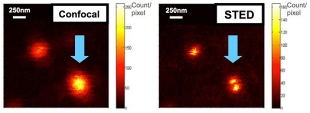
Fig 1: Scanning images of the microscope PSF in confocal mode (left) and STED mode (right) probed by 40nm fluorescent beads. STED clearly resolves the two adjacent fluorescent beads.
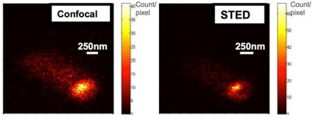
Fig. 2: Preliminary fluorescence images of PopZ-GFP proteins in a live Caulobacter crescentus cell. The autofluorescent background marks the approximate outline of the cell. The STED image (right) exhibits finer features than the confocal counterpart (left).
Andrew Olson
Neuroscience
Project: Neuroscience Microscopy Service 3D Image Analysis Resource
Other Key Personnel
Gary Steinberg, M.D., Ph.D., Director, SINTN
Stephen J. Smith, Ph.D., Professor of Molecular and Cellular Physiology
Jon Mulholland, Director, Cell Sciences Imaging Facility
Summary
Many modern microscopy techniques require capital-intensive equipment (e.g., two photon microscopy, single photon confocal microscopy), along with expertise in its proper use. Recognizing this, the Stanford Institute for Neuro-Innovation and Translational Neuroscience (SINTN) has recently established a microscopy core facility, the Neuroscience Microscopy Service (NMS; began operations in August, 2008). The NMS provides a centralized resource for two-photon microscopy, confocal microscopy, and Array Tomography [1] data acquisition. During the initial period of operation of the NMS, we have identified 3D image analysis as a key need among the neuroscience faculty that we believe the NMS is ideally positioned to address. With funding from this grant, we propose to create the nucleus for a 3D Image Analysis Center, by purchasing a high-end image analysis workstation and 3D analysis software.
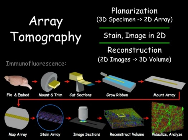
Visual overview of the steps involved in Array Tomography. (Smith lab)
Yuzuru Takashima
Electrical Engineering
Project: Development of high throughput and high resolution NSOM probe.
Other Key Personnel
Yao-Te Cheng, Ph.D. Student, Materials Sci. and Eng.
Summary
The research aims to provide a C-Aperture Nano Tip (CAN-Tip) NSOM imaging probe, having high power throughput (>x104), high resolution (<λ/50) and low background noise, for use by the nano-imaging research community at Stanford University.
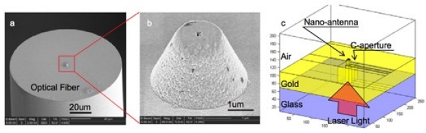
NSOM probe with C-shaped aperture fabricated on optical fiber (a), (b) and schematic of proposed NSOM probe having C-shaped aperture with nano optical antenna (c).
Philip Yang
Cardiovascular Medicine
Project: Molecular MRI for Differentiation, Imaging, and Selection of Cardiac Progenitor Cells
Other Key Personnel
Dwight Nishimura, PhD, Professor, Electrical Engineering
Renee Reijo-Pera, PhD, Professor, Stem Cell Institute
Thomas Quertermous, MD, Professor, Cardiovascular Medicine
Robert Robbins, MD, Professor, Cardiothoracic Surgery
Summary
To date, no reliable method for cardiac transdifferentiation of pluripotent embryonic stem cells (ESCs) has emerged [1-2]. In order to address this issue, this proposal will outline a combinatorial differentiation protocol, which utilizes a novel exogenous protein, apelin, and a MRI reporter gene (RG). First, our differentiation protocol employing apelin generated cardiac progenitor cells marked by increased early expression of cardiac development genes and contractile cardiomyocytes in mouse ESCs. This protocol will be optimized by the addition of known mesodermal differentiation factors at temporally specific time points. Second, our lentiviral RG construct has demonstrated a robust genomic integration and expression of fusion protein consisting of two cell surface tags and luciferase in both mouse and human ESCs. This construct will be modified to integrate cardiac-specific transcription factors to direct the apelin-prepped mesodermal cells into cardiac lineage. This proposal is designed to induce robust cardiac differentiation, image the differentiation process, and select the cardiac progenitor cells.
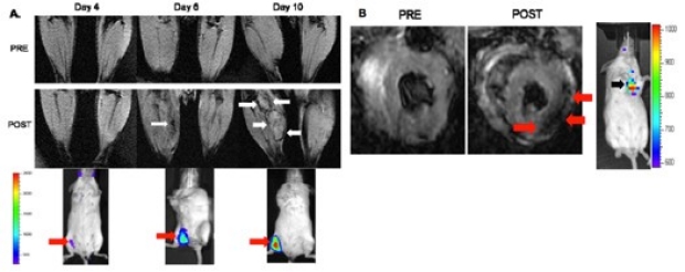
In vivo molecular MRI of hESCs-RG transplanted into mouse hind limbs and myocardium. A. Longitudinal in vivo MRI and concurrent BLI of mouse hind limbs after transplantation of 1X10⁶ hESC-RG in the right hind limb and of the non-transduced mESCs in the contralateral hind limb. Following intravenous administration of microbeads, viability signal was first noted from hESCs-RG on day 6 and increased by day 10 (white arrows). BLI validates MRI signal on the corresponding right hind limb (red arrows). B. Similarly, in vivo molecular MRI of hESCs-RG transplanted into the mouse myocardium following intravenous administration of microbeads is shown (red arrows). PRE refers to pre-injection of microbeads and POST to post-injection of microbeads. The MRI signal is validated by a positive BLI signal.
Guy Ziv
Biochemistry
Project: Studying actin-based motility in 3D using light-field microscopy
Other Key Personnel
Julie Theriot, Associate Professor, Biochemistry Department, Stanford University
Marc Levoy, Professor, Computer Science Department, Stanford University
Matt Footer, Research Associate, Biochemistry Department, Stanford University
Summary
Specific Aims of Research:
1. Construct a light-field microscope capable of simultaneous tracking of multiple objects in 3D
2. Adapt and develop novel algorithms based on computational photography for 3D particle tracking
in light-field microscopy.
3. Study the kinetics of actin-polymerization driven beads in 2D and 3D.
4. Compare experimental observations to models of actin-based motility
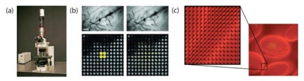
(a) Image of the prototype light-field microscope. The microlens array is marked with a red circle. (b) Two synthetically focused images of Golgi-stain slice of rat brain. Upper images show the computationally focused image, lower images show which pixels of the 4D light-field data were used to calculate the intensity at the center pixel of each image. (c) Sample of fixed mouse intestine villus infected with Listeria. Zooming in on the overhead view in the right, one can see the numerous subimages produced by each microlens. Within each of these subimages, the position defines the angle of incoming rays. Images reproduced from [28] and the Stanford light-field micrscope project website http://www-graphics.stanford.edu/projects/lfmicroscope/