2011 CBIS Seed Grant Recipients
Molecular & Cellular Biology
Project: New Laboratory Component of MCP 222 - Biological Light Microscopy
Summary
[pending]
Gregory B. Rieker, PhD
Mechanical Engineering
Project: Medical Imaging Isotope Production Using a Novel Compact Plasma Accelerator
Gregory B. Rieker, PhD
Mechanical Engineering
Project: Medical Imaging Isotope Production Using a Novel Compact Plasma Accelerator
Summary
The goal of this project is to demonstrate production of the PET isotope 13N using a compact plasma accelerator technology developed by researchers in the Stanford Plasma Physics Laboratory (SPPL). Current medical accelerators are complex and expensive, which limits the widespread applications of isotopes to the few that have sufficient half-lives to be generated in large, off-site facilities and transported to the patient. Researchers in the SPPL are developing a compact plasma-based accelerator technology with significantly reduced complexity and cost compared with traditional ion-based accelerators. This project is also meant to foster collaboration between the SPPL and the Molecular Imaging Program at Stanford (MIPS). The 13N produced in the SPPL will be rapidly transported to the Radiochemistry Facility of the MIPS program for accurate yield measurements.
Other Key Personnel
Flavio Poehlmann, PhD, Mark Cappelli, PhD
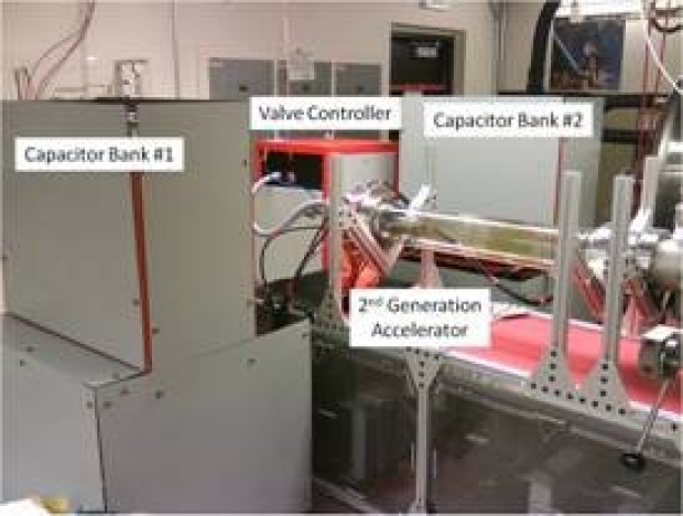
Compact plasma accelerator system
Dimitre Hristov, PhD, MCCPM
Radiation Oncology
Project: Magnetic Resonance Imaging of Radiation-Activated Alginate Nanoparticle Drug/Sensitizer Delivery
Summary
The effectiveness of chemotherapeutic agents or radio-sensitizers administered concurrently with radiation therapy is limited by their systemic toxicity. Hence there is need for the development of targeted oncologic image-guided drug delivery approaches for concentrated and controlled release in tumors in order to minimize drug-induced adverse effects without compromising tumor control.
Our goal is to investigate the feasibility of a novel image-guided drug delivery approach that employs liquid-core nanocapsules releasing their core drug and MR contrast content in response to ionizing radiation. As conformal radiation therapy techniques focus radiation on the tumor, this approach will lead to tumor-targeted drug delivery with reduced exposure of healthy tissues to the drug.
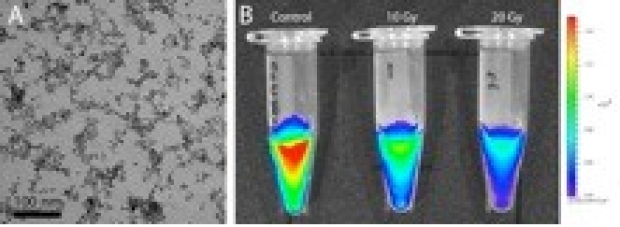
Nanoparticle characterization and alginate radiolysis (A) Transmission Electron Microscopy of Super-paramagnetic Iron Oxide Nanoparticles and (B) Alginate nanoparticles displaying core content release after irradiation.
Geoffrey A. Kerchner, MD, PhD
Neurology
Project: Microstructural Correlates of Motor Deficits in Parkinson Disease with Ultra-High Field 7-Tesla MRI
Summary
Parkinson’s disease involves the accumulation of microscopic protein deposits in brain neurons, and the eventual atrophy of selected populations of neurons in the midbrain and elsewhere. These changes are evident at autopsy, but not on conventional clinical MRI. This work aims to take advantage of the potential for ultra-high field 7-Tesla MRI to produce images of high spatial resolution to visualize structural changes in midbrain structures in patients with Parkinson’s disease, and to correlate these changes with clinical metrics of motor function. This effort may lead to the development of an imaging biomarker capable of distinguishing Parkinson’s disease from related disorders, and to track the progress of the disease over time.
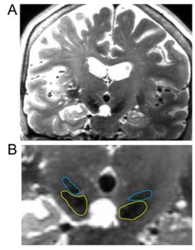
Figure. 7T 3D T2-weighted CUBE imaging, shown in the coronal plane and windowed so as to emphasize midbrain anatomy. This image was obtained in a volunteer patient with Alzheimer’s disease. (A) Entire coronal slice through the midbrain. Changes in tissue contrast across the slice reflect radiofrequency inhomogeneities, with no correction applied to this raw image. (B) Same image, zoomed to the midbrain. The STN is outlined in blue, and the SN in yellow. Parameters: 17x17 cm FOV, 224x224 matrix, 0.8 mm slice thickness, TE 70 mx, cardiac gates with 5 RR (TR ~5000 ms), 256 slices, scan time ~5 min.
Yaping Joyce Liao, MD, PhD
Ophthamology
Project: In Vivo Imaging of the Mouse Optic Nerve in Aging and in Experimental Ischemic Optic Neuropathy
Summary
Axons are the information highways that connect the 100 billion neurons and form the 100 trillion synapses in the human brain. These brain connections are lost as part of normal aging. Because the optic nerve contains the only axons that can be imaged in high resolution in the living human, changes in the optic nerve may be the early signs of not only optic nerve diseases but also central nervous system conditions. Our goals include: 1) to study the changes in axons and axonal transport in normal aging and following experimental anterior ischemic optic neuropathy (AION), the most common acute optic neuropathy in older adults, and 2) the use of intravitreal and systemic pharmacophore trk receptor agonists to bypass the axon-transport dependency of neurotrophic support to treat experimental AION. Our findings will provide valuable insight on the axonal changes in normal aging and alterations following injury that may underlie selective vulnerability in older mammals.
Other Key Personnel
Frank Longo, MD PhD
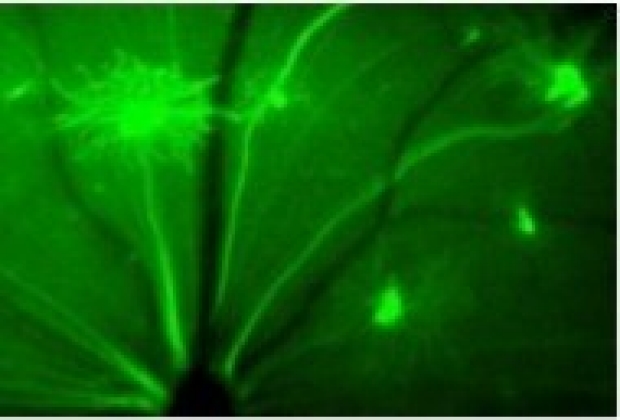
In vivo imaging of retinal ganglion cell axons, dendrites and cell bodies in the Thy1-YFP mice.
Saikat Pal, PhD
Bioengineering
Project: A Novel Imaging Technique to Acquire Joint Kinematics and Stress for Diagnosis of Knee Osteoarthritis
Summary
The purpose of this study is to acquire accurate three-dimensional kinematics and cartilage-bone stress at the knee joint under weightbearing conditions in healthy controls and symptomatic subjects. We are interested in understanding the mechanisms of joint pain and cartilage degeneration by quantifying differences in kinematic patterns between controls and symptomatic subjects, potentially leading to abnormal loading, elevated cartilage-bone stress, and degeneration of the knee joint.
Other Key Personnel
Rebecca Fahrig, PhD, Garry Gold, MD, Scott Delp, PhD, Jang-Hwan Choi, MS, Thor Besier, PhD
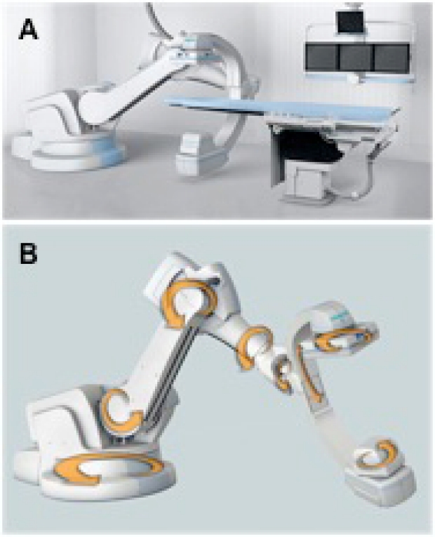
Figure 1: (a) Typical clincal installation of the zeego system with the arm in "side" position, and (B) the robot has 8 degrees of positioning freedom including rotation of detector and x-ray tube.
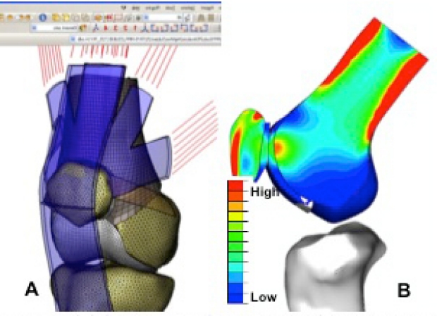
Figure 2: (A) Subject-specific FE model of the knee, and (B) predicted cartilage-bone stress during a weightbearing squat.
Cara H. Haney, PhD
Biology
Project: Using Array Tomography to Study Coordination of Extracellular Matrix and Cell Behavior in Plant Development
Summary
Plant development is strikingly different from animal development starting with this plain fact: from birth to death, plant cells exist in a living box, the cell wall. This dynamic organelle is composed of complex carbohydrates and proteins. The role of the extracellular matrix is acknowledged to be important in all multicellular organisms; but particularly in plants, cell wall activities are the major driving force in morphogenetic decisions and reactions. Thus, the cell wall is not just a consequence of plant cell differentiation, but also a player in determining future cell function and fate. We will probe sections of a plant meristem using antibodies specific for diverse wall components, and analyze these by array tomography. This will establish for the first time in any plant system a 3-dimensional, chemically analyzed map of how cell type and wall composition correspond with developmental change.
Other Key Personnel
David Ehrhardt, Ph.D. Staff Scientist, Carnegie Institution
Elizabeth Wiltshire, graduate student, Biology department
Sharon Long, PhD
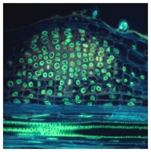
This developing root nodule is beginning to differentiate distinct layers with specific cell characteristics. Antibodies that recognize specific complex carbohydrates will be used to probe cell wall variations during development.
Manjula Kurella Tamura, MD, MPH
Nephrology
Project: Relation Between Chronic Kidney Disease, Hypertension Control and Brain Perfusion
Summary
Our overarching goal is to characterize the physiological mechanisms that underlie the relationship between chronic kidney disease, hypertension control, and small vessel ischemic disease in the brain. In this seed grant we will conduct preliminary studies to measure brain perfusion using Arterial Spin Labeling (ASL) in persons with chronic kidney disease.